


Views: 0 Author: Site Editor Publish Time: 2026-08-07 Origin: Site


While monolithic zirconia is marketed as one of the most durable restorative materials in modern dentistry, its ultimate clinical performance is strictly governed by preparation geometry and material thickness. Dental professionals frequently encounter catastrophic fractures, margin chipping, or retention failures. These issues are rarely due to defective material. They typically stem from inadequate wall thickness, improper yttria selection, or insufficient cement space during the preparation phase.
Mitigating these risks requires a precise understanding of how structural dimensions interact with material science. This guide establishes evidence-based parameters for wall thickness, evaluating how different zirconia generations and restoration types dictate preparation requirements. By mastering these dimensions, you ensure predictable seating, optimal stress distribution, and long-term clinical success without compromising the underlying tooth structure.
Yttria Content Defines Thickness Limits: Stronger 3Y-zirconia requires a minimum of 0.4 mm (anterior) to 0.5 mm (posterior), whereas highly translucent 4Y and 5Y variants demand 0.8 mm to 1.0 mm for structural integrity.
Axial Wall Compensation: When occlusal clearance is clinically limited, maintaining a thicker axial wall can compensate for stress distribution and prevent flexural fatigue.
Retention vs. Thickness Trade-offs: Over-tapering axial walls to achieve recommended thickness compromises the resistance form, leading to premature debonding regardless of material strength.
Digital Workflow Tolerances: Modern milling requires precise cement space calculations; insufficient space prevents complete seating, artificially introducing stress concentrations at the margins.
The Microleakage Risk: Suboptimal wall thickness and improper margin design lead to open margins and flexural gaps, allowing saliva and bacteria to seep under the restoration, resulting in secondary caries and cement dissolution.
Zirconia relies on phase transformation toughening to resist fracture. When a micro-crack begins to form under heavy occlusal load, the material undergoes a localized volume expansion at the crack tip. This expansion compresses the defect and halts its progression through the ceramic matrix. However, this mechanism requires adequate material volume. Sufficient thickness allows the restoration to absorb heavy forces without propagating structural defects. If the walls are too thin, the material lacks the necessary bulk to execute this phase transformation, leading to catastrophic failure under stress.
Clinical experience shows that fractures rarely happen on day one. They occur after thousands of cyclic loading events. A well-prepared tooth provides the necessary foundation for the ceramic to function as designed. When you compromise on reduction, you force the material to absorb forces it cannot handle, bypassing its inherent toughening mechanisms.
The structural integrity of a zirconia crown depends heavily on its yttria concentration. Increasing yttria content improves translucency by stabilizing the cubic phase, making the material look more like natural enamel. Unfortunately, this aesthetic improvement significantly reduces fracture toughness. There is a direct correlation between yttria content and preparation requirements. Lower flexural strength materials like 4Y and 5Y mandate a proportional increase in minimum wall thickness. You must prepare the tooth deeper to achieve the same clinical survivability as traditional 3Y materials.
Zirconia Generation | Yttria Content | Flexural Strength | Clinical Indication |
|---|---|---|---|
3Y-TZP | 3 mol% | 1000 - 1200 MPa | Posterior restorations, high-stress areas, bruxers. |
4Y-PSZ | 4 mol% | 700 - 900 MPa | Anterior restorations, premolars with adequate clearance. |
5Y-PSZ | 5 mol% | 500 - 700 MPa | High-aesthetic anterior cases, strict thickness compliance needed. |
Occlusal forces do not remain trapped in the restorative material. They transfer through the crown down into the underlying dentin. Uniform thickness prevents localized stress pooling. When a preparation features sharp internal line angles or uneven reduction, force concentrates at the thinnest points. A smooth, uniform preparation allows masticatory forces to dissipate evenly across the entire abutment. This mechanical harmony prevents flexural fatigue and protects the underlying tooth structure.
To achieve optimal stress distribution, follow these clinical steps during preparation:
Use a depth-cutting bur to establish uniform reduction guidelines across the occlusal surface.
Reduce the axial walls using a flat-end taper bur, maintaining a consistent angle.
Round all internal line angles with a fine diamond bur to eliminate stress concentrators.
Verify clearance using a flexible clearance tab in all excursive movements.
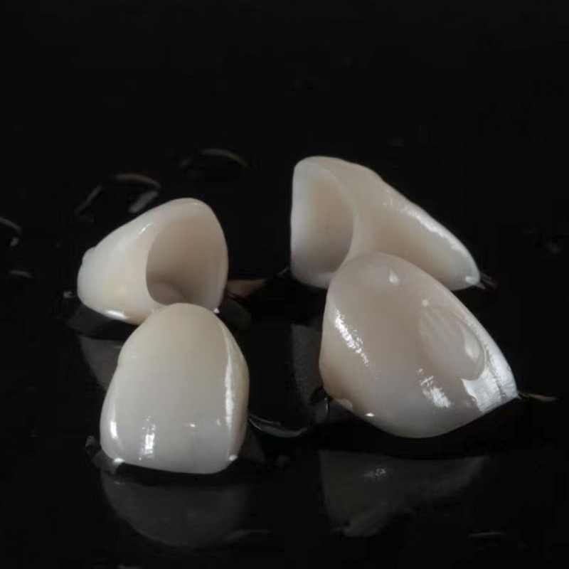
Anterior restorations require precise reduction to balance aesthetics and strength. For high-strength 3Y zirconia, you need a minimum wall thickness of 0.3 mm to 0.4 mm. If you select high-translucency 4Y or 5Y variants, the minimum thickness jumps to 0.8 mm. Lingual clearance is equally critical. You must provide enough incisal edge reduction to prevent shear fractures during protrusive movements. Inadequate lingual reduction forces the laboratory to thin the material precisely where anterior guidance exerts the most lateral stress.
When preparing anterior teeth, the path of insertion must align with the adjacent teeth while respecting the pulp chamber. Aggressive reduction for 5Y materials on mandibular incisors often risks pulpal exposure. In these cases, switching to a stronger 3Y or 4Y material allows for a more conservative preparation while maintaining structural integrity.
Posterior teeth endure heavy masticatory forces. You must establish a baseline occlusal thickness of at least 0.5 mm for 3Y materials. For 4Y or 5Y variants, you need a minimum of 1.0 mm of occlusal clearance. Functional cusp reduction is non-negotiable. You must bevel the functional cusp to provide uniform bulk over the primary stress-bearing areas. Avoid sharp internal line angles at all costs. Sharp angles act as stress concentrators that initiate internal cracks within the ceramic framework.
Evaluating posterior clearance requires more than just having the patient bite down in centric occlusion. You must check clearance during lateral and protrusive excursions. A preparation that looks adequate in centric occlusion may severely lack clearance when the patient grinds laterally, leading to premature fracture of the restoration.
Zirconia possesses exceptionally high edge strength compared to traditional glass ceramics. This property allows for thinner margins, such as a feather-edge or a light chamfer. However, you must evaluate the threshold where margin thinness transitions from clinically acceptable to a milling liability. If the margin is too thin, the milling burs will cause micro-chipping during manufacturing. This clinical risk is severe. Chipped margins lead to open margins. Open margins allow saliva and bacteria to infiltrate the cement space, causing biological failure and secondary decay.
Laboratories typically recommend a heavy chamfer or a modified shoulder for optimal milling results. These margin designs provide enough bulk for the milling burs to cut cleanly without vibrating and chipping the fragile pre-sintered ceramic. A well-defined margin also makes it significantly easier to read the digital scan and design a precise fit.
An implant zirconia crown operates under different biomechanical rules than a tooth-supported restoration. Implants lack a periodontal ligament (PDL). The PDL normally acts as a shock absorber during mastication. Without it, the crown absorbs direct, rigid impact forces. This requires stricter adherence to minimum occlusal thickness guidelines. You must ensure robust thickness at the abutment-crown interface to prevent catastrophic material failure under heavy loading.
When designing for implants, the screw access hole introduces a weak point in the ceramic structure. The material surrounding this access hole must be thick enough to withstand torque during insertion and occlusal forces during function. Titanium bases are often used to reinforce this connection, transferring stress away from the ceramic and into the implant fixture.
Conservative dentistry often favors partial coverage. When preparing a zirconia inlay onlay, the design principles shift. Unlike full-coverage crowns, inlays and onlays rely heavily on internal cavity dimensions. You must maintain a minimum isthmus width of 1.5 mm to 2.0 mm. Depth requirements are equally strict. Adequate bulk prevents the restoration from fracturing under lateral excursive forces. Smooth transitions and rounded internal angles are mandatory to prevent wedging forces from splitting the natural tooth.
Preparation for partial coverage requires divergent internal walls. Any undercuts will prevent the rigid ceramic from seating. Block out undercuts with composite resin before taking the final impression or digital scan. This ensures a passive fit and reduces internal stress on the ceramic during cementation.
Modern digital dentistry relies heavily on scanner-first workflows. When designing a custom zirconia crown, lab technicians use CAD software to identify thin spots in real-time. The software generates digital thickness maps. Clinicians must evaluate these maps carefully before approving the final design. If the software highlights areas below the minimum thickness threshold, you must either adjust the preparation clinically or accept a compromised, over-contoured final restoration.
Communication with the laboratory is critical. Provide detailed instructions regarding the desired yttria generation and the specific occlusal scheme. If the preparation is borderline thin, instruct the lab to prioritize structural integrity over ideal anatomy. A slightly flatter occlusal table is preferable to a restoration that fractures within the first year of service.
Clinicians frequently face the reality of short clinical crowns. A short preparation height severely compromises the resistance form of the final restoration. If you aggressively reduce the occlusal surface to achieve ideal material thickness, you may lose the axial wall height needed for retention. The mitigation strategy is straightforward. You can increase the axial wall thickness to compensate for compromised occlusal thickness. This maintains overall structural rigidity without over-tapering the preparation.
In cases of extreme wear, consider increasing the vertical dimension of occlusion (VDO) before preparing the teeth. This provides the necessary restorative space without requiring aggressive reduction of already compromised tooth structure. Crown lengthening surgery is another viable option to gain axial wall height and improve retention.
Inadequate cement gap parameters in CAD/CAM design lead to hydraulic pressure during cementation. This pressure prevents complete crown seating. The balance of cement thickness is delicate. Insufficient cement space raises the occlusion and leaves margins open. Conversely, excessive cement thickness degrades retention. A thick cement layer is prone to wash-out when exposed to oral fluids. Incomplete seating leaves margins open to microleakage. Saliva and bacteria seep in, dissolving the cement and compromising the underlying tooth structure.
To ensure proper seating, follow these cementation protocols:
Clean the intaglio surface of the restoration with a zirconia cleaner to remove salivary phosphates.
Isolate the preparation to prevent moisture contamination during cementation.
Apply firm, even pressure during the initial set of the cement to overcome hydraulic resistance.
Remove excess cement promptly before it fully hardens to prevent marginal irritation.
There is a dangerous trade-off between aggressive reduction and the loss of axial wall parallelism. Clinicians sometimes over-taper the preparation to achieve ideal material thickness quickly. This combination of a short crown height and over-tapered walls compounds the risk of mechanical debonding. You must maintain a 6- to 10-degree total occlusal convergence. This specific taper ensures mechanical retention alongside material strength, preventing the crown from dislodging during sticky food consumption.
If a preparation becomes over-tapered, you must rely on adhesive resin cements rather than conventional luting agents. Resin cements provide chemical bonding that compensates for the lack of mechanical retention. However, this requires strict isolation and precise bonding protocols to achieve a durable result.
Audit your current preparation protocols and standardize bur selections to ensure predictable reduction depths across all cases.
Collaborate closely with your digital dental laboratory to verify thickness and cement space parameters prior to milling.
Select the appropriate yttria generation based on the available restorative space and the functional demands of the specific tooth.
Implement strict isolation and cleaning protocols during cementation to maximize bond strength and prevent microleakage.
A: For traditional 3Y zirconia, the absolute minimum occlusal thickness is 0.5 mm. However, 1.0 mm is highly preferred for optimal stress distribution. If you are using more translucent 4Y or 5Y variants, you must maintain at least 1.0 mm of thickness to prevent fracture under heavy masticatory forces.
A: Higher yttria content (4Y/5Y) increases the material's translucency but significantly decreases its flexural strength. Because the material is inherently weaker, your preparations must be 0.3 mm to 0.5 mm thicker than those required for traditional, high-strength 3Y zirconia.
A: Yes. High-strength zirconia can tolerate feather-edge margins due to its exceptionally high edge strength. However, your milling equipment must be perfectly calibrated. Poor calibration will cause micro-chipping at the thin edges, resulting in open margin gaps during seating.
A: Thin axial walls lack the volume needed to absorb occlusal loads. This leads to flexural fatigue under lateral forces. Over time, this flexing increases the risk of vertical fractures, margin deformation, and ultimate failure of the underlying cement bond.
A: If the cement space is too tight, hydraulic pressure prevents the crown from seating fully. This creates unsupported gaps and high-stress contact points. Conversely, excessive cement thickness compromises mechanical retention and increases the risk of margin microleakage and cement wash-out.
A: Yes. Implants lack a periodontal ligament, meaning they cannot absorb shock like natural teeth. Implant crowns absorb direct, rigid force. This makes strict adherence to minimum occlusal thickness guidelines absolutely critical to prevent catastrophic material failure.
A: Inadequate reduction at the margin forces the lab to create either over-contoured crowns or dangerously thin margins that chip during milling. Both scenarios result in open margins. Saliva and bacteria easily seep into these gaps, causing secondary decay and cement dissolution.


