


Views: 0 Author: Site Editor Publish Time: 2026-08-22 Origin: Site


Zirconia is widely celebrated for its extreme durability and esthetic potential in restorative dentistry. However, this same rigidity presents an unforgiving clinical challenge. Achieving and maintaining uniform contacts requires absolute precision. Both occlusal and interproximal areas demand perfection. Uneven contacts carry severe clinical risks. They can trigger rapid opposing dentition wear. They also cause catastrophic micro-fractures. Furthermore, persistent implant screw loosening frequently occurs. When these failures happen, clinicians face frustrated patients. They also suffer costly clinical remakes. Resolving these complications requires completely moving away from analog guesswork. Instead, maintaining uniform contacts demands a stringent digital bridge scan workflow. You must pair this digital foundation alongside precise material selection. You also need meticulous post-mill polishing protocols. Read on to discover how mastering digital precision safeguards long-term restorative success. We will explore the exact biomechanics involved. We will overcome common milling limitations. Finally, we will detail essential clinical adjustments. Our goal is ensuring predictable, stress-free delivery appointments.
Digital Predictability: Intraoral scanning and CAD/CAM workflows eliminate the dimensional shrinkage associated with traditional impression materials, ensuring highly accurate interproximal and occlusal mapping.
Material Unforgivingness: Unlike acrylic or composite, a custom zirconia bridge will not "wear in." Uniform contacts must be engineered prior to final seating to prevent opposing enamel damage.
Milling & Sintering Science: Precision relies on calculating the exact shrinkage rate of zirconia during the sintering phase to maintain contact integrity.
Adjustment Protocols: Proper polishing—not just glazing—is required post-adjustment to maintain long-term contact uniformity without abrasive wear.
Evaluating the stakes of asymmetrical contacts reveals serious clinical consequences. Premature contacts quickly disrupt masticatory function. Open margins invite bacterial microleakage around the abutments. Both issues severely compromise the immediate success of any restoration. Biomechanics play a critical role here. Localized high spots on rigid zirconia act as destructive force multipliers. They transfer excessive occlusal forces directly to the abutments. This pressure eventually reaches the underlying alveolar bone. Consequently, biological complications accelerate rapidly. You often see early signs of peri-implantitis or crestal bone resorption.
The business impact is equally severe. High spots demand heavy chairside adjustments. Time spent grinding down a custom implant bridge significantly reduces case profitability. A routine thirty-minute delivery can easily become a stressful ninety-minute ordeal. You lose valuable scheduling blocks. Furthermore, aggressive chairside grinding introduces invisible micro-cracks into the material structure. If clinicians fail to cool the zirconia properly during adjustment, heat builds up. Catastrophic fractures often follow shortly after the patient leaves.
Evidence-oriented realities strongly support these claims. Many dentists mistakenly assume material defects cause restorations to break. However, clinical data tells a very different story. An estimated majority of zirconia bridge failures actually stem from unbalanced occlusion. Fractures or opposing wear rarely originate from inherent flaws in the manufactured puck. Instead, they result directly from poor force distribution.
Common Mistake: Relying solely on articulating paper marks without asking the patient about proprioceptive feedback. Zirconia's rigidity means even microscopic high spots feel massive to the patient's neuromuscular system.
Analog impression distortions often ruin well-planned cases. Polyvinyl siloxane and polyether materials naturally shrink over time. Moisture and temperature changes warp physical models during transit. You can bypass these dimensional errors entirely. Simply rely on a digitally native process. Digital files maintain permanent dimensional stability. They capture soft tissue and abutment positions without physical distortion. Scanning wands map the exact emergence profiles flawlessly.
Virtual articulation offers another massive advantage. Advanced CAD software simulates dynamic mandibular movements effortlessly. Lab technicians use these tools to design uniform functional contacts. They evaluate the restoration in static centric relation. They also map excursive movements. You no longer guess where lateral interferences might occur. Software highlights collisions using color-coded heat maps.
Interproximal calculations become incredibly precise within a digital environment. Digital design tools allow technicians to adjust contact broadness accurately. They modify tightness down to a single micron level. This level of precision ensures a highly predictable, passive fit. The restoration slides perfectly against adjacent natural teeth or abutments. It prevents the dreaded "black triangle" effect between teeth.
Data verification acts as a final risk-mitigation step. Top-tier labs print resin try-ins before milling final restorations. We highly recommend utilizing these inexpensive prototypes. Clinicians evaluate the resin try-in directly in the mouth. You can verify occlusal schemes and interproximal tightness safely. You can make adjustments chairside and rescan the modified prototype. Once approved, the lab mills the final zirconia block using verified data.
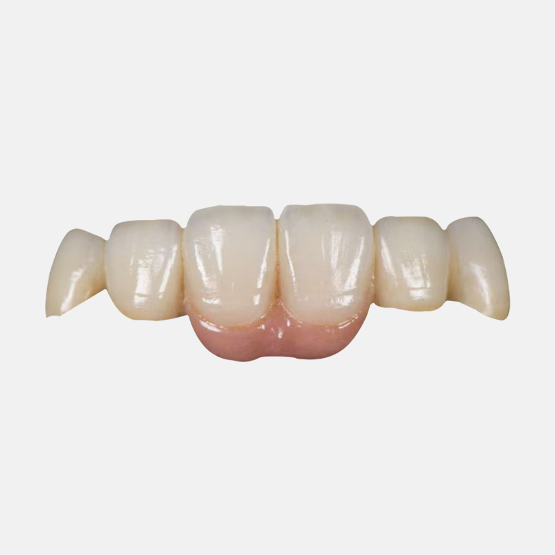
Managing contacts on a single quadrant requires standard restorative care. However, addressing a full arch zirconia bridge presents a distinct, larger-scale problem. Entirely different biomechanical rules apply to full mouth rehabilitations. You are essentially rebuilding the patient's entire functional system from scratch.
Bilateral balanced occlusion becomes an absolute necessity. Masticatory forces must distribute evenly across the entire arch simultaneously. Proper distribution prevents dangerous cantilever fractures. It also prevents facial support collapse over time. Patients rely on this equilibrium for comfortable chewing. Without balance, one side bears the brunt of the biting force. This imbalance eventually loosens the retaining screws.
Material dynamics dictate a very cautious approach. Monolithic zirconia boasts an impressive flexural strength. It typically ranges from 900 to 1200 MPa. Because of this massive strength, the material will never yield. It does not compress under pressure like natural ligaments do. Therefore, you must engineer exact opposing contact distribution. Any localized pressure points will destroy opposing natural enamel rapidly. The zirconia will simply obliterate softer materials.
We must acknowledge a transparent assumption about full-arch cases. Many clinicians assume the bridge itself holds the sole responsibility for occlusion. This assumption is completely false. Achieving uniform contacts on full arches often requires additional work. You usually need meticulous equilibration of the opposing dentition. Clinicians must adjust natural teeth or existing restorations. They must harmonize the entire stomatognathic system before seating the final prosthesis.
Best Practices for Full Arch Occlusion:
Map excursive pathways carefully using digital articulation software.
Eliminate severe canine guidance interferences beforehand.
Equilibrate opposing natural teeth to flatten excessively steep cusps.
Utilize rigid resin try-ins to verify bilateral stability clinically.
We must evaluate the highly technical laboratory process. Unprocessed zirconia remains relatively soft in its green state. It undergoes a massive transformation during the high-heat sintering process. Specifically, zirconia shrinks approximately 20 to 25 percent inside the furnace. This extreme shrinkage presents a serious dimensional hurdle for contact accuracy.
Advanced CAM software calibration solves this issue. High-tier dental labs rely on sophisticated nesting software. These programs calculate exact shrinkage rates based on specific puck batches. They scale the digital design up perfectly before milling begins. This precise calibration ensures the planned contacts match the finalized custom zirconia bridge exactly. If the algorithm misses by a fraction of a percent, contacts open up or bind.
Milling hardware also imposes strict physical limitations. High-speed milling burs sometimes struggle to reach tight spaces. They cannot always carve sharp interproximal embrasures perfectly. Round burs naturally leave slightly rounded internal geometries. Therefore, skilled manual refinement remains necessary before the final bake. Expert technicians meticulously refine the green-state zirconia under magnification.
They ensure contacts remain broad and uniform. Pinpointed contacts cause severe food impaction. They also create isolated instability. Proper hand-refinement eliminates these micro-errors entirely.
Table: Processing Variables for Contact Predictability
Processing Phase | Primary Objective | Risk if Mishandled |
|---|---|---|
CAM Nesting | Calculate 20-25% volume shrinkage accurately | Open interproximal margins or altered occlusion |
Machine Milling | Translate digital design into green-state block | Rounded embrasures due to bur size limitations |
Manual Refinement | Shape broad, uniform interproximal contact areas | Pinpointed contacts leading to localized pressure |
Clinical implementation introduces a major risk factor regarding surface treatment. Clinicians frequently adjust contacts chairside to perfect the bite. However, this necessary grinding instantly removes the protective surface glaze. If left unpolished, the underlying rough zirconia acts like coarse sandpaper. It brutally grinds against opposing natural teeth during every chewing cycle.
Implementing a strict polishing protocol is completely non-negotiable. You cannot simply smooth the surface using standard finishing burs. Instead, you must use specialized diamond-impregnated polishers. These tools systematically reduce surface roughness at a microscopic level. They effectively restore a high-gloss, ultra-smooth finish. This vital step protects opposing enamel from severe, irreversible abrasion.
We must also confront a stark clinical reality about glazes. Surface glazes look beautiful upon delivery. They provide a highly esthetic shine initially. However, they routinely wear off over 6 to 12 months. This degradation occurs most rapidly in high-contact occlusal areas. Glaze cannot withstand constant masticatory friction.
Therefore, uniform, highly polished monolithic surfaces remain superior. They offer the only verifiable way to maintain safe, long-term uniform contacts. When the glaze inevitably fades, the polished core material remains smooth.
Recommended Polishing Protocol Sequence:
Adjust high spots using a fine-grit diamond bur under copious water cooling.
Apply a medium-grit diamond-impregnated rubber wheel to smooth the adjusted zone.
Use a fine-grit polishing point to begin restoring the surface luster.
Finish with a high-shine zirconia polishing paste and a bristle brush.
Securing a successful restorative outcome involves far more than material selection alone. A durable restoration relies heavily on the precision of data captured. It equally depends on your chosen laboratory's advanced CAD/CAM expertise. Analog methods simply cannot compete with modern computational accuracy. Digital ecosystems eliminate the guesswork.
Clinicians must carefully establish strict decision criteria for lab partners. You should evaluate labs based on their digital workflow integration. Prioritize technicians who mandate resin try-in protocols for complex cases. Furthermore, demand clear guidelines regarding material-specific shrinkage compensation. These vital factors dictate the final seating accuracy. They determine whether you spend five minutes or fifty minutes adjusting the bite.
Take action to elevate your restorative predictability today. We strongly encourage adopting a fully digital scanning protocol immediately. Abandon outdated, unstable impression materials. Partner with a specialized restorative lab for your upcoming bridge cases. Embracing these advanced methodologies ensures your restorations function flawlessly. They will provide patients with years of comfortable, balanced function.
A: Minor discrepancies frequently occur due to subtle jaw position changes during intraoral scanning. Slight sintering variations in the lab can also alter dimensions. Even minor shifts create noticeable high spots because zirconia does not compress. Careful chairside adjustment and thorough repolishing are strictly required to resolve this interference.
A: Zirconia itself resists wear exceptionally well and rarely changes shape. However, uniform contacts can shift over time. This shifting usually happens if opposing natural teeth wear down. It also occurs if patients experience underlying implant bone loss, which subtly alters the restorative biomechanics.
A: Digital workflows drastically reduce chair time and eliminate many micro-errors. However, they cannot account for everything. Human biology constantly shifts. The periodontal ligament (PDL) flexes naturally during function. Because of these biological variables, minor clinical refinements remain the standard of care for achieving perfect equilibrium.


