


Views: 0 Author: Site Editor Publish Time: 2026-10-14 Origin: Site


Have you ever designed a flawless digital crown, only to watch it fail spectacularly in the mouth? Imagine the clinical frustration. The patient is in the chair. The soft tissue is beautifully prepped. Yet, the definitive zirconia restoration simply refuses to seat. This nightmare scenario usually traces back to a microscopic scanning error that compromised your entire digital foundation.
The shift from traditional elastomeric impressions to digital workflows has revolutionized modern implantology. At the very center of this transformation is the Scanning Body. Clinicians frequently refer to it as a scan flag or scan peg. This small device acts as the critical physical-to-digital bridge. Without absolute precision at this stage, even the most advanced intraoral scanners cannot prevent misfits. You will inevitably face costly laboratory remakes and compromised patient outcomes.
In this comprehensive guide, we will explore why component accuracy dictates the trajectory of the entire restorative phase. We will also analyze the common reasons digital impressions fail and outline best practices for preventing costly misfits.
Precision Equals Passive Fit: The primary role of a scan flag is to transfer the exact three-dimensional position and angulation of the implant to the CAD software. Even microscopic inaccuracies lead to poorly fitting prosthetics.
Material and Design Matter: The geometry, material composition, and surface finish significantly impact the intraoral scanner's ability to capture data accurately without light distortion.
Human Error is a Major Factor: Improper seating due to tissue interference, blood or saliva contamination, and erratic scanning paths are the leading clinical causes of scan failures.
Quality Accessories Save Money: Investing in premium components reduces chair time, eliminates expensive lab remakes, and ensures long-term biological and mechanical implant success.
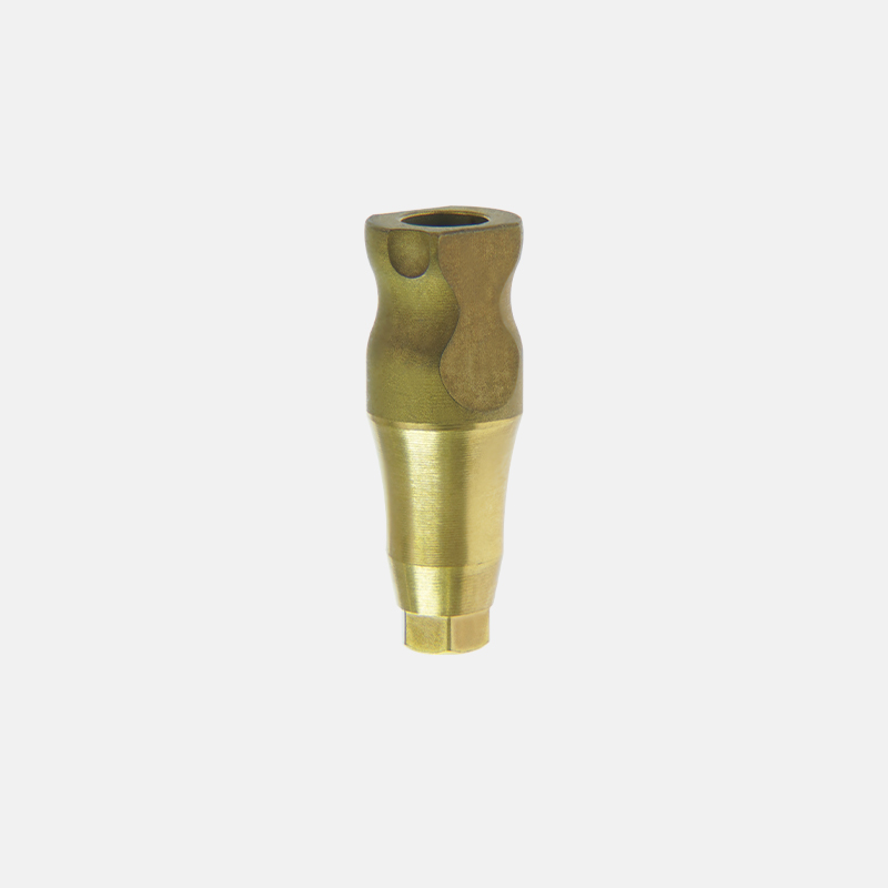
A scan peg is a precisely manufactured component that screws directly into the implant fixture or a multi-unit abutment. Its sole purpose is to represent the exact location, depth, and rotational orientation of the implant within the virtual environment. When captured by an intraoral scanner, the design software reads the specific geometry of the component. The software then superimposes a digital replica of the implant from its pre-loaded digital library.
To fully grasp how this device functions, we must break down its physical anatomy. Each segment serves a distinct, non-negotiable purpose in the digital workflow.
Anatomical Part | Primary Function | Clinical Importance |
|---|---|---|
The Base | Interfaces directly with the implant connection such as internal hex or conical. | Must be machined to exact tolerances to ensure a flush, accurate seat without micro-gaps. |
The Body | The vertical, scanable surface captured by the intraoral scanner's cameras. | Requires a matte finish to prevent light reflection and optical scattering during the scan. |
The Flat Surface Bevel | An asymmetrical flat plane located on the upper portion of the body. | Indicates the exact rotational timing, also known as anti-rotation, of the implant to the CAD software. |
While it may appear to be a simple piece of metal or plastic, this component is the foundational data point for the entire CAD/CAM process. As a highly critical Digital Dental Accessory, its precision dictates the success of every subsequent step. If the initial data captured from this tool is flawed, the digital design, the milling process, and the final delivery will all be built upon a fundamentally incorrect foundation.
In the realm of digital dentistry, errors do not isolate themselves. They compound. A minor dimensional discrepancy at the scanning phase creates a ripple effect that ultimately compromises the final prosthetic. Understanding the severe consequences of these inaccuracies highlights exactly why precision remains non-negotiable.
In implant prosthetics, achieving a passive fit is the ultimate clinical objective. Unlike natural teeth, which are suspended in the periodontal ligament and possess a slight degree of mobility, dental implants are rigidly osseointegrated into the jawbone. They have absolutely zero tolerance for misfit.
If the Implant Scan Body is misaligned by even a few microns due to poor manufacturing tolerances or improper seating, the digital model will misrepresent the fixture's true position. Consequently, the milled crown or titanium framework will not seat passively. Forcing a poorly fitting restoration into place introduces severe mechanical stress directly to the implant components and the surrounding crestal bone.
The downstream consequences of an inaccurate scan extend far beyond an initial seating struggle. When a restoration lacks a passive fit, microscopic gaps form at the implant-abutment interface. From a biological standpoint, these micro-gaps act as a protected harbor for bacterial colonization. The resulting bacterial leakage predictably leads to localized inflammation, peri-implant mucositis, and eventually peri-implantitis. This is a primary driver of marginal bone loss and complete implant failure.
Furthermore, the continuous stress of a non-passive restoration leads to repeated screw loosening, screw fracture, or even catastrophic sheer fracture of the implant fixture itself. Absolute accuracy at the initial impression phase serves as the first line of defense against these devastating long-term complications.
Time remains one of the most valuable assets in any busy dental practice. When an inaccurate digital file results in a crown that refuses to fit, the immediate consequence is lost chair time. The clinician is forced to remove the restoration, retake the digital impression, and schedule the frustrated patient for another visit. In addition, laboratory remakes incur significant financial penalties. Utilizing highly precise components streamlines the workflow, making delivery appointments entirely predictable while maximizing practice profitability.
Even when utilizing state-of-the-art intraoral scanners, digital impressions can and do fail. Interestingly, these failures are rarely the fault of the hardware itself. Instead, they are most often attributed to human error, anatomical challenges, or compromised clinical accessories.
The single most prevalent cause of scan failure is the incomplete seating of the component. In many clinical scenarios, overgrown gingival tissue, tight interproximal contacts from adjacent teeth, or residual crestal bone spicules physically prevent the base from engaging fully with the implant connection. If the base is elevated by just a fraction of a millimeter, the software will render the implant higher than its true depth. This results in a restoration that suffers from hyper-occlusion or fails to seat entirely. Clinicians must perform adequate bone profiling and meticulous tissue management to guarantee a clear path of insertion.
Many practices utilize reusable models to reduce overhead. However, repeated clinical use and sterilization take a severe toll. Polyether ether ketone materials, while excellent for optical capture due to their non-reflective nature, are susceptible to thermal degradation. Repeated autoclave cycles can warp the plastic, subtly altering the geometry of the critical flat bevel. In contrast, titanium versions are highly durable but can become scratched over time. Scratches on a metal surface create erratic light reflections, confusing the scanner's cameras and leading to distorted 3D meshes. Regular inspection of your Intraoral Scan Body inventory is absolutely mandatory.
Intraoral scanners operate by capturing thousands of overlapping images and stitching them together to form a cohesive 3D model. Operator technique heavily influences the software's ability to stitch these frames accurately. Moving the wand too rapidly, hovering at steep angles, or failing to capture the distinct flat plane head-on can cause the software to guess the geometry. This leads to severe alignment errors. A methodical, steady scanning path is required to feed the software clean, unambiguous data.
The digital dentistry landscape is shifting rapidly. Current market analyses reveal a distinct evolution in restorative protocols. As artificial intelligence becomes deeply embedded in CAD software for automated margin detection and mesh alignment, the software's tolerance for ambiguous optical data is shrinking. Software algorithms now demand absolute clarity.
Consequently, the industry is moving decisively toward highly integrated digital ecosystems. Clinicians increasingly recognize that relying on degraded or generic scan flags introduces unacceptable variables. The standard of care is shifting toward premium components engineered in tandem with robust digital libraries. This ensures zero discrepancy between the physical impression and the virtual model.
The dental market offers a vast array of aftermarket options. Each boasts distinct characteristics. The material composition and geometric design directly influence how easily and accurately the optical sensors can capture the required data.
Clinicians generally choose between three main material classifications. To understand the operational impact, we must look at the specific engineering parameters.
Material Type | Optical Scanability | Autoclave Durability | Clinical Recommendation |
|---|---|---|---|
All-PEEK Polymer | Excellent matte finish without scanning spray. | Low to Medium. Prone to thermal warping. | Best suited for strict single-use protocols. |
Grade 5 Titanium | Naturally reflective. Requires matte spray or sandblasting. | Extremely High. Resists deformation. | Ideal for high-wear clinical environments. |
Premium Hybrid Design | Flawless upper capture via matte PEEK. | High base precision via Titanium interface. | The modern gold standard for digital implantology. |
Specifically, premium hybrid components combine a highly precise Grade 5 Titanium base with a matte PEEK upper body. High-end manufacturing maintains tolerances as tight as ±5 microns at the connection interface. This level of precision eliminates rotational play and ensures the digital mesh aligns perfectly with the CAD library.
Beyond material, the physical shape matters immensely. Scanners rely on distinct geometric features to orient the digital model in a 3D space. Designs with well-defined, sharp flat planes are significantly easier for the software algorithms to recognize than those with overly rounded or ambiguous features. Additionally, the surface finish must remain entirely matte. Any glossy or reflective surface will cause the scanner's projected light to scatter, resulting in data voids in the final mesh.
To achieve predictable, passive-fitting restorations, clinicians must adopt a standardized, uncompromising protocol for digital impressions. Implementing the following best practices will drastically reduce the margin of error.
Intraoral scanners are optical devices. They cannot see through fluids. Before inserting the component, the implant connection must be thoroughly irrigated and dried. The presence of blood, saliva, or crevicular fluid inside the internal hex can physically prevent complete seating. Furthermore, if saliva coats the outer surface during the scan, it creates a reflective sheen that refracts light and distorts the data. Proper isolation using cotton rolls, retraction cords, or high-volume suction is a critical preparatory step.
Assuming a component is fully seated based solely on tactile feedback is a dangerous clinical gamble. Soft tissue impingement can often mimic the satisfying click of a fully seated component. Taking a periapical radiograph to verify complete seating is a mandatory, non-negotiable step before ever picking up the scanner. The X-ray provides objective, visual proof that the base is perfectly flush with the implant platform.
A strategic scanning path ensures the software captures the geometry efficiently without triggering stitching errors. Begin by scanning the occlusal surface, ensuring the flat bevel is captured head-on with high data density. Next, slowly roll the scanner to the buccal and lingual aspects while maintaining a steady pace. Finally, ensure the camera captures the emergence profile. This transition area where the component meets the soft tissue is vital for designing the emergence profile of the final crown.
While scanning a single unit is relatively straightforward, full-arch implant rehabilitations introduce complex biomechanical and digital challenges that push optical scanners to their absolute limits.
In full-arch cases, minor inaccuracies compound exponentially. If a single post is distorted by just 20 microns, that error is magnified as the scanner moves across the featureless edentulous ridge. This leads to severe cross-arch distortion. A titanium framework milled from distorted data will not seat passively across all multi-unit abutments. This often requires the clinician to section and solder the framework, completely defeating the purpose of a digital workflow.
To combat cross-arch distortion, the industry has developed advanced solutions. Horizontal designs feature geometric shapes spanning laterally across the arch, providing the scanner with continuous data points to reduce stitching errors. Furthermore, photogrammetry has emerged as a superior technology for complex cases. Instead of relying on a continuous video scan, photogrammetry uses specialized fiducial markers and stereoscopic cameras to capture the exact spatial relationship of the implants in a single flash. This bypasses the inherent compounding errors of traditional intraoral scanning.
Selecting the appropriate components is a critical business and clinical decision. The right tools will streamline your workflow, while inferior aftermarket products will introduce endless clinical frustration.
The physical piece of hardware is entirely useless without its corresponding digital file. Before purchasing, you must verify that the physical geometry exactly matches the CAD library utilized by your dental laboratory, such as Exocad, 3Shape, or Dental Wings. If the physical shape does not perfectly align with the digital library file, the software will superimpose the implant incorrectly, leading to immediate restorative failure.
Clinicians must weigh the operational costs of single-use versus reusable options. Single-use PEEK models guarantee pristine geometry for every scan, eliminating the risk of autoclave distortion, but they increase per-patient overhead. Reusable titanium or hybrid options require a higher initial investment but offer long-term durability, provided they are meticulously maintained and discarded at the first sign of surface scratching.
Precision machining is the absolute cornerstone of digital dentistry. The tolerances required for a base to engage an implant connection are measured in microns. Purchasing cheap, off-brand components often results in poorly machined interfaces that wobble or fail to seat accurately. Investing in premium, precision-engineered products is a safeguard for your clinical reputation. Sourcing a high-quality Implant Accessory ensures the tight manufacturing tolerances required for predictable, passive-fitting restorations. Partnering with reputable manufacturers guarantees that the physical hardware and the digital software work in perfect harmony.
The scan peg may be one of the smallest tools in your surgical kit, but it is undeniably one of the most critical. It dictates the accuracy of the entire digital implant workflow from start to finish. Cutting corners on these essential components causes massive time losses, financial drains, and can ultimately damage patient trust when a final restoration fails to fit.
Audit your current digital dental workflow today. Discard worn-out or heavily autoclaved polymers immediately, and transition to reliable, precision-milled components. By investing in premium digital accessories from LEZY, you protect your clinical outcomes, streamline your chair time, and guarantee a passive fit for every patient who walks through your door. Visit our catalog today to find the exact specifications tailored to your digital library.
Yes, many models are specifically engineered for multiple uses, but they require rigorous inspection protocols. PEEK materials are susceptible to structural deformation and dimensional changes after repeated exposure to the high heat and pressure of autoclave sterilization cycles. Conversely, titanium models are highly durable but can suffer from surface scratching, which may cause optical scattering during the digital impression. Clinicians must strictly adhere to the manufacturer's specified limits on reuse cycles and discard components at the first sign of visible wear.
Scanning difficulties generally stem from three primary clinical factors. First, high light reflection can confuse the optical sensors, particularly if you are using an untreated shiny titanium surface without applying a scanning spray. Second, the presence of biological fluids, such as saliva, blood, or crevicular fluid, alters the surface geometry and refracts the scanner's light. Third, inadequate ambient lighting or poor isolation can hinder data acquisition. Ensuring the surgical site is thoroughly isolated and perfectly dry is the most effective troubleshooting step.
Yes, taking a periapical radiograph is universally considered a mandatory clinical step. It is the only definitive, objective method to confirm that the component is fully and passively seated into the implant's internal connection. Soft tissue impingement or unseen bone spicules can easily prevent complete seating, leading to a distorted digital impression and a misfitting final restoration.
An implant-level component connects directly to the internal or external interface of the implant fixture itself, capturing its precise spatial orientation within the jawbone. In contrast, an abutment-level component attaches to an intermediate structure, such as a multi-unit abutment. Abutment-level scanning is predominantly utilized in complex, full-arch restorations to correct extreme angulations and simplify the final prosthetic framework.
The CAD software relies on a specific, pre-installed digital library provided by the manufacturer. The unique physical geometry of the hardware corresponds exactly to a digital mesh file within that library. When the software recognizes the scanned shape, it superimposes the exact implant interface, determining its depth, angulation, and rotational timing within the 3D digital model.
Reusable components must be cleaned manually or in an ultrasonic cleaner using enzymatic solutions to remove all biological debris before sterilization. Avoid highly abrasive brushes that could scratch the surface. Once cleaned and thoroughly dried, they should be placed in a sterilization pouch and autoclaved according to the manufacturer's specific temperature and cycle parameters. Never exceed the recommended number of sterilization cycles for polymer-based items.
Not necessarily. Single crowns typically utilize implant-level components that capture the exact anti-rotation features of the implant fixture. For full-arch cases, clinicians generally use abutment-level components that seat on multi-unit abutments. Furthermore, advanced full-arch cases often require specialized horizontal designs or photogrammetry flags to provide the scanner with additional reference points, drastically reducing cross-arch distortion.


