


Views: 0 Author: Site Editor Publish Time: 2026-08-28 Origin: Site


Clinical frustration hits hard when a perfectly milled implant-supported restoration suffers from decementation or delamination. You deal with the hidden costs of chairside remakes, disrupted schedules, and unhappy patients. These failures turn routine restorative procedures into clinical liabilities. The shift from traditional castable UCLA abutments toward pre-machined, screw-retained hybrid crowns streamlined digital workflows and improved marginal adaptation. However, this shift introduced a distinct biomechanical challenge. The physical dimensions of the abutment interface—specifically vertical height—often get compromised due to interarch space limitations or reliance on standardized inventory.
When vertical space is tight, you are forced to use shorter abutments. Reducing vertical wall height directly compromises the resistance form. This leaves the cement layer vulnerable to catastrophic shear forces during mastication. Understanding the biomechanical relationship between abutment height and structural retention dictates long-term restorative success. We will evaluate how to select correct abutment dimensions, optimize surface protocols, and identify exactly when to pivot to alternative restorative solutions.
The 3.5mm Threshold: In-vitro evidence consistently demonstrates that abutment heights exceeding 3.5mm significantly reduce the risk of crown debonding, regardless of the restorative material used.
Surface Area vs. Resistance Form: Vertical height exponentially increases the surface area for cementation while providing critical resistance against lateral occlusal forces.
Surface Preparation Multiplies Retention: Mechanical retention relies on more than just height; a taller base does not guarantee success if surface prep is ignored. Proper surface treatment (e.g., sandblasting) can increase bond strength up to fivefold.
Standard vs. Custom: Standardized Ti-bases are highly efficient, but specific clinical scenarios (extreme angulation, offset implant placement, deep emergence profiles) strictly require a custom abutment.
Establishing a predictable baseline for implant-supported restorations demands meeting strict clinical success criteria. A successful restoration must exhibit flawless marginal integrity to prevent bacterial microleakage. It needs occlusal stability to distribute masticatory forces evenly across the arch. It requires soft tissue health to maintain crestal bone levels and support the gingival architecture. Above all, it requires absolute retention. If the overlying crown detaches from the substructure, all other clinical parameters fail instantly. The foundation of this retention lies in the geometric design and structural integrity of the titanium base abutment.
The physics of debonding are driven by the complex forces exerted during the chewing cycle. Mastication does not merely apply vertical compressive loads. It introduces severe lateral and shear forces across the occlusal table. When a patient grinds or chews tough foods, the resulting off-axis forces attempt to tip the crown off its foundation. If the vertical walls of the abutment are insufficient to block this tipping motion, the entire load transfers directly to the cement layer. Dental cements perform exceptionally well under compression but fail rapidly under shear stress. Once the shear forces exceed the cohesive strength of the cement, micro-fractures propagate through the luting agent. This inevitably leads to complete crown delamination.
This biomechanical vulnerability is severely exacerbated by offset or off-axis implant placement. In ideal scenarios, the surgeon positions the implant fixture directly beneath the central fossa of the planned restoration. This allows forces to travel straight down the long axis of the implant body. However, anatomical limitations—such as a narrow alveolar ridge or proximity to the maxillary sinus—often force surgeons to place implants at an angle or slightly off-center. Offset placement creates a cantilever effect. It acts as a lever arm that multiplies the lateral forces applied to the restorative interface. Under these conditions, the shear stress on the cement layer increases dramatically. The vertical height of the abutment becomes the primary mechanical resistance to counteract this lever arm.
The titanium base serves as the central load-bearing hub between the osseointegrated implant fixture and the rigid restorative material. It must absorb, distribute, and transfer occlusal loads without yielding. Because it acts as the primary interface, its geometric proportions dictate the long-term survival of the restoration. A base that is too narrow or too short fails to provide the required surface area for chemical bonding. It lacks the physical bulk needed to resist lateral tipping. Evaluating the exact dimensions of this interface is the first step in preventing restorative failure.
To understand the failure mechanics, we must look at the specific stress distribution patterns. When a lateral force hits the buccal cusp of a mandibular molar implant crown, the crown attempts to rotate lingually. The buccal margin goes into tension, while the lingual margin goes into compression. The vertical wall on the lingual side of the abutment must physically block this rotation. If the wall is too short, the crown rotates past the abutment, tearing the cement bond on the buccal side. This is why height is not just about surface area; it is about creating a physical barricade against rotation.
The mathematical relationship between vertical wall height and resistance form is a fundamental principle of restorative dentistry. Resistance form relies on the presence of opposing vertical walls that physically block the displacement of the crown when lateral forces are applied. As the height of the abutment wall increases, the pivot point of the crown shifts. This reduces the tangent angle of displacement. Taller walls effectively intercept the internal surface of the crown before the cement layer can be stressed to the point of failure. This physical blocking mechanism prevents the crown from tipping under off-axis loading.
There is a direct and exponential correlation between abutment height and total cementable surface area. Dental cements require adequate surface area to establish a durable micromechanical and chemical bond. By increasing the vertical height from 3.0mm to 4.0mm, the total circumferential surface area available for cementation expands significantly. This increased area distributes the functional loads over a wider region. It reduces the localized stress concentrations that typically initiate cement fatigue.
Utilizing short abutments introduces severe biomechanical risks. These risks are primarily characterized by specific failure modes such as zirconia delamination and rapid cement fatigue. When an abutment measures less than 3.0mm in height, it lacks the necessary vertical geometry to provide adequate resistance form. Consequently, the rigid overlying material—particularly monolithic zirconia—transfers nearly all occlusal stress directly into the thin cement space. Because zirconia does not flex under load, the underlying cement absorbs the entirety of the shock. This leads to rapid micro-cracking and eventual cohesive failure.
Despite these risks, the clinical reality of posterior restorations often dictates the use of shorter bases. In the molar regions, interocclusal space is frequently limited due to supereruption of opposing teeth or naturally tight vertical dimensions. You are forced to choose between reducing the thickness of the restorative material (risking crown fracture) or utilizing a shorter abutment (risking decementation). When forced into this compromise, you must aggressively manage the biomechanical trade-offs through enhanced surface treatments and strict adhesive protocols. The mechanical retention provided by the short wall is inherently deficient.
Current in-vitro consensus and extensive clinical testing indicate that a minimum height of 3.5mm to 4.0mm is required for the predictable, long-term retention of hybrid-abutment-crowns. This specific range is considered the biomechanical sweet spot. At 3.5mm and above, the vertical walls provide sufficient resistance form to counteract lateral forces. This drastically reduces the reliance on the cement's shear strength. Achieving this height threshold significantly improves retention universally, regardless of whether the overlying crown is milled from rigid monolithic zirconia or slightly more elastic lithium disilicate.
This 3.5mm to 4.0mm height also strikes the optimal balance between abutment retention and restorative material thickness. It leaves adequate space above the abutment to ensure the overlying crown meets the manufacturer's minimum thickness requirements for structural durability. Simultaneously, it maintains an optimal cement space geometry. This allows for complete seating of the restoration without hydraulic pressure buildup, ensuring a passive fit and a uniform cement layer.
Failure Modes by Abutment Height
Vertical Height | Primary Failure Mode | Clinical Recommendation |
|---|---|---|
< 3.0mm | Rapid cement shear failure, crown delamination | Avoid if possible. Mandates aggressive sandblasting and MDP primers. |
3.0mm - 3.4mm | Moderate cement fatigue under heavy lateral loads | Acceptable for anterior teeth or premolars with flat occlusal schemes. |
3.5mm - 4.0mm | Highly stable. Rare cohesive cement failure. | Ideal target for all posterior and anterior restorations. |
> 4.0mm | Restorative material fracture (if interarch space is limited) | Ensure minimum 1.5mm occlusal clearance remains for the ceramic. |
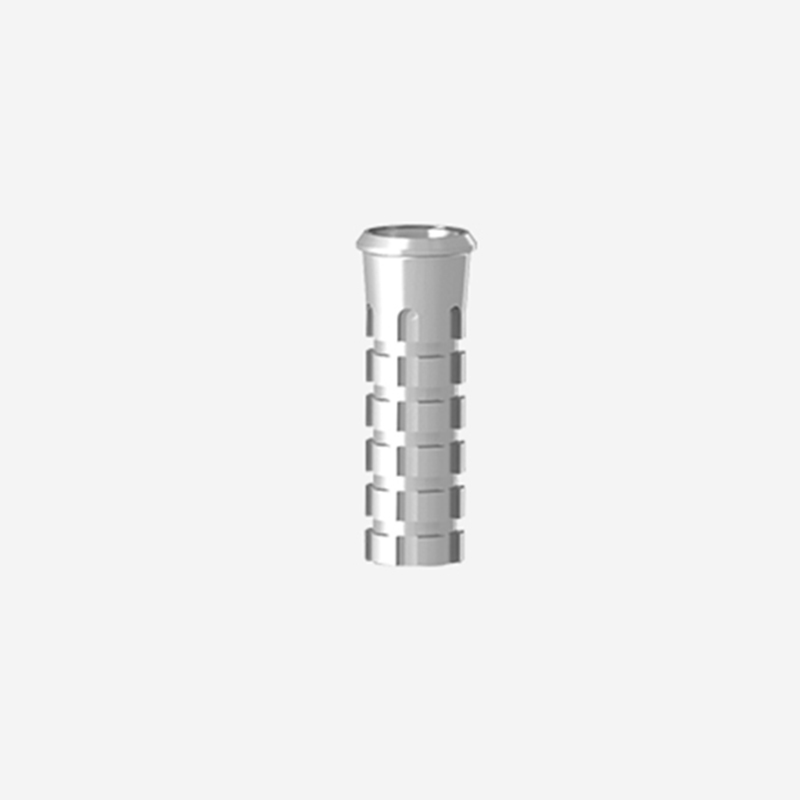
Standardized titanium bases offer workflow efficiency, cost-effectiveness, and inventory scalability. For routine, straightforward cases with ideal implant placement, parallel trajectories, and adequate interarch space, standard bases provide a highly predictable solution. Dental laboratories maintain a streamlined inventory of standard heights and emergence profiles. This allows for rapid CAD/CAM design and turnaround times. The pre-machined implant connection ensures absolute precision at the fixture level, minimizing the risk of micro-gap formation and bacterial infiltration.
The limitations of standardized components become apparent in complex clinical scenarios. Standard bases feature fixed emergence profiles that may not adequately support the soft tissue architecture in highly scalloped anterior regions. They utilize standardized margin heights that cannot adapt to uneven crestal bone levels or varying gingival depths. Most critically, standard bases lack the ability to correct severe implant angulations. When an implant is placed at a trajectory that deviates significantly from the planned restorative axis, a straight standard base forces the screw access hole through the facial or incisal edge of the restoration. This compromises both aesthetics and structural integrity.
Specific clinical parameters completely disqualify the use of a standard base and mandate the fabrication of a custom abutment. Any implant placement requiring an angulation correction greater than 15 degrees typically exceeds the design limits of standard components. Highly scalloped anterior tissue architectures require customized subgingival contours to support the papillae and create a natural emergence profile. Excessively deep implant placement also necessitates a custom solution to elevate the restorative margin to a cleanable, supragingival or slightly subgingival level. This prevents the catastrophic complication of retained subgingival cement.
Many top-tier clinicians prefer a custom abutment by default for any case outside of the posterior mandible. They argue that the ability to control the exact emergence profile, margin placement, and vertical wall height yields superior long-term tissue stability and retention. While the initial laboratory cost of a patient-specific abutment is higher, the long-term return on investment is substantial. Investing in customized geometry upfront drastically reduces the risk of soft tissue recession, peri-implantitis, and the costly, time-consuming process of replacing a failed standard restoration.
When evaluating the cost-to-benefit ratio, you must factor in chair time. Seating a crown on a standard base that requires heavy occlusal adjustment because the angulation was slightly off takes 30 minutes. Seating a crown on a custom abutment designed to perfectly parallel the adjacent teeth takes 5 minutes. The laboratory bill might be higher for the custom part, but the clinical overhead drops significantly. Furthermore, the custom abutment allows the technician to place the cement margin exactly 0.5mm subgingivally across the entire circumference of the tooth, regardless of how uneven the bone crest is. A standard base forces a flat margin, which might be 2mm subgingival on the mesial and supragingival on the distal.
Clinical Indications for Abutment Selection
Clinical Parameter | Standard Ti-Base | Custom Abutment |
|---|---|---|
Implant Angulation | Ideal trajectory (< 15° deviation) | Severe deviation (> 15° correction needed) |
Tissue Architecture | Flat, thick biotype (Posterior) | Highly scalloped, thin biotype (Anterior) |
Implant Depth | Standard crestal placement | Excessively deep subcrestal placement |
Interarch Space | Adequate vertical clearance (> 7mm) | Compromised vertical clearance |
Emergence Profile | Symmetrical, standard healing | Asymmetrical, requires specific tissue support |
While vertical height provides the macroscopic resistance form, the microscopic surface topography of the abutment dictates the ultimate tensile strength of the cement bond. Machined titanium is inherently smooth. It offers virtually no micromechanical retention for luting agents. To overcome this, you must mechanically alter the surface. Sandblasting the titanium surface with aluminum oxide creates a highly textured, micro-retentive landscape. This process exponentially increases the available surface area at a microscopic level. It allows the cement to flow into the irregularities and interlock upon curing.
The dependency between abutment height and surface preparation cannot be overstated. Industry data consistently demonstrates that proper surface treatment can increase retention strength by up to five times compared to untreated, machined titanium bases. This creates a specific clinical dynamic: a shorter, properly sandblasted base can frequently outperform a taller, untreated base in shear bond strength tests. Maximizing retention requires treating height and surface topography as interdependent variables. Relying solely on a 4.0mm height without executing strict surface abrasion protocols will still result in premature debonding.
The chemical interface between the titanium and the restorative material is heavily influenced by cement selection. Resin cements are universally preferred for cementing an implant crown titanium base due to their superior compressive strength, low solubility, and ability to form chemical bonds with specialized primers. Resin-modified glass ionomer (RMGI) cements lack the sheer bond strength required to secure rigid ceramics to non-porous titanium. They are useful for natural teeth due to moisture tolerance, but they fail on titanium interfaces.
Successful extraoral cementation demands strict implementation protocols to prevent contamination. The titanium surface must be thoroughly cleaned to remove all residual alumina particles and handling oils. Following cleaning, the application of an MDP-containing primer (10-Methacryloyloxydecyl dihydrogen phosphate) is mandatory. The MDP monomer chemically bonds to the titanium oxide layer on the abutment and the oxide networks in zirconia restorations. This creates a robust, durable chemical linkage. Complete isolation during this extraoral process is essential. Even microscopic salivary or sebaceous contamination will instantly neutralize the chemical bonding sites, leading to inevitable failure.
Different restorative materials exhibit distinct behavioral characteristics when subjected to occlusal stress. This directly impacts how they interact with varying heights of titanium bases. Monolithic zirconia possesses an exceptionally high modulus of elasticity, making it incredibly rigid. When lateral forces are applied to a zirconia crown, the material does not absorb or dissipate the energy through flexure. Instead, it transfers the entire load directly to the cement interface. If the underlying abutment walls are short, this rigid transfer of force rapidly shatters the cement layer.
Materials like lithium disilicate possess a slightly lower modulus of elasticity. This allows for a microscopic degree of flexure under load. While this flexure can help dissipate some stress, it introduces the risk of material fracture if the underlying abutment does not provide adequate uniform support. Achieving the 3.5mm height threshold improves retention for all materials, but rigid materials like zirconia are particularly unforgiving of short abutment walls. When utilizing zirconia on compromised vertical heights, the reliance on flawless surface treatment and MDP chemical bonding becomes absolute.
To guarantee maximum retention, dental laboratories must follow a rigid extraoral cementation protocol. Skipping a single step compromises the entire assembly.
Protect the implant connection interface using a dedicated analog or polishing protector. Do not allow sandblasting media to touch the internal connection.
Sandblast the retention walls of the titanium base using 50-micron aluminum oxide at 2 bar (30 psi) pressure from a distance of 10mm for exactly 10 seconds.
Clean the abutment in an ultrasonic bath with 99% isopropyl alcohol for 3 minutes to remove all dust and oils.
Apply an MDP-containing metal primer to the sandblasted titanium surface. Allow it to react for 20 seconds, then gently air-dry until the solvent evaporates.
Sandblast the intaglio surface of the zirconia crown (at 1 bar pressure) or etch the lithium disilicate crown with hydrofluoric acid.
Apply the corresponding ceramic primer to the intaglio surface of the crown.
Extrude dual-cure resin cement into the crown, seat the abutment firmly, and maintain continuous pressure while tack-curing the margins.
Remove excess cement immediately, apply glycerin gel to the margins to prevent an oxygen inhibition layer, and complete the final light cure.
The foundation of a perfectly fitting implant restoration begins with flawless data acquisition. The primary function of the dental scan body is to accurately transfer the precise 3D position, trajectory, and rotational timing of the implant fixture into the CAD software. This specialized component acts as a digital beacon. It allows the intraoral scanner to map the exact spatial coordinates of the implant connection relative to the adjacent dentition and opposing arch.
Beyond capturing the implant's position, the scan data maps the surrounding soft tissue depth and the available interocclusal clearance. These two spatial metrics directly dictate the maximum allowable vertical height for the titanium base. If the scan reveals a shallow tissue profile and limited interarch space, the CAD technician is immediately alerted to the vertical constraints. Accurate capture of this data ensures that the subsequent design phase is grounded in physical reality. It prevents the selection of an abutment height that would violate the required restorative material thickness or impinge on the opposing occlusion.
Once the digital impression is imported, the CAD technician must select the appropriate component library within the design software. This selection process involves evaluating the available abutment heights against the mapped interocclusal space. The software aligns the digital replica of the abutment with the scanned position. This allows the technician to visualize the exact relationship between the top of the abutment and the opposing cusp tips.
Setting the correct design parameters ensures the structural integrity of the final restoration. The technician must establish an optimal cement gap—typically between 30 to 50 microns. This allows for adequate cement flow without creating a loose fit that compromises resistance form. The software enforces minimum material thickness rules based on the selected restorative material. If a taller abutment encroaches on this minimum thickness, the software highlights the collision. This forces the technician to adjust the emergence profile, reduce the abutment height, or switch to a custom solution to safely accommodate the necessary restorative bulk.
The digital workflow also allows for the precise calculation of the emergence angle. If the implant is placed deeply, the software can calculate the exact angle required to bring the restorative margin from the implant platform to the gingival crest. If this angle exceeds 30 degrees, the soft tissue will likely recede due to excessive pressure. The technician can visualize this in the CAD software and adjust the abutment design accordingly. Standard bases do not offer this flexibility, forcing the tissue to adapt to a pre-machined shape, which often leads to ischemia and subsequent bone loss.
CAD Design Parameters for Hybrid Crowns
Design Parameter | Recommended Setting | Clinical Rationale |
|---|---|---|
Radial Cement Gap | 30 - 40 microns | Provides enough space for resin cement without losing frictional resistance. |
Occlusal Cement Gap | 50 - 60 microns | Allows hydraulic pressure to escape during seating, preventing incomplete seating. |
Minimum Zirconia Thickness | 1.0mm (Axial), 1.5mm (Occlusal) | Prevents catastrophic fracture of the ceramic under heavy masticatory loads. |
Margin Thickness | 0.2mm - 0.3mm | Ensures the milling bur can accurately cut the margin without chipping the ceramic. |
To immediately improve clinical outcomes and reduce the incidence of decementation, practices and laboratories should take the following actions:
Audit your current restorative inventory to ensure all standard bases meet the minimum 3.5mm height requirement whenever anatomically possible.
Standardize your extraoral cementation protocols by mandating 50-micron aluminum oxide sandblasting and MDP primer application for every single case.
Establish strict communication thresholds with your laboratory partners to automatically transition to custom solutions when implant angulation exceeds 15 degrees.
Implement rigorous intraoral scanning techniques to accurately capture soft tissue depth and interocclusal clearance, providing the lab with exact spatial data.
A: The clinical and biomechanical threshold is between 3.5mm and 4.0mm. Heights within this range provide sufficient vertical wall geometry to create resistance form, which physically prevents the overlying crown from tipping under lateral masticatory forces and significantly reduces shear stress on the cement layer.
A: Yes, sandblasting is mandatory. It creates a micro-retentive surface topography that can increase cement bond strength by up to five times compared to untreated titanium. Height alone is insufficient; without micromechanical retention and proper chemical priming, even tall abutments are susceptible to decementation.
A: A custom solution is mandatory when dealing with severe implant angulations (requiring >15-degree correction), excessively deep subcrestal placement, highly scalloped anterior tissue that demands specific support, or when offset placement requires unique structural reinforcement that standard bases cannot provide.
A: A titanium base features a pre-machined, highly precise implant connection and is designed for extraoral cementation of a CAD/CAM milled crown. A traditional UCLA abutment relies on a castable plastic sleeve that is waxed and cast in metal, which often results in a less precise implant-to-abutment interface.
A: While limited interarch space often forces the use of bases under 3.0mm, it significantly increases the risk of delamination due to high occlusal forces. To mitigate this, you must maximize surface treatments (sandblasting, MDP primers) or pivot to custom abutments to optimize the available retention.
A: The scan body accurately transfers the 3D position, trajectory, and surrounding soft tissue depth to the CAD software. This precise digital mapping dictates the available interocclusal clearance and margin depth, directly determining the maximum allowable vertical height for the abutment.


