


Views: 0 Author: Site Editor Publish Time: 2026-08-22 Origin: Site


Placing implants at steep angles allows clinicians to avoid anatomical structures like the maxillary sinus. However, it transfers significant complexity directly to the restorative phase. Without precise data transfer, angled implants lead to compromised screw access channels, non-passive fits, and biomechanical failure over time. Transitioning from surgical placement to the final implant supported bridge requires strict evaluation. You must analyze how spatial data is captured, interpreted, and applied by the dental laboratory. Small deviations multiply across the arch, putting restorative success at risk. Designing a predictable, long-lasting prosthesis requires specific angulation metrics and a clear choice between abutment systems. You will learn how modern data-capture workflows minimize clinical distortion. We will explore exactly what metrics prevent hardware fatigue and guarantee a flawless structural fit for your patients.
Data specificity prevents failure: Accurate capture of the implant trajectory, depth, and restorative space is non-negotiable for achieving a passive fit.
Workflow dictates accuracy: Evaluating photogrammetry versus traditional intraoral scanning (IOS) depends on the span of the bridge and the severity of the implant angles.
System selection hinges on angulation limits: Angles exceeding 30 degrees typically mandate a multi unit abutment bridge to correct trajectories, whereas minor discrepancies may allow for a custom abutment bridge.
Biomechanical risks are quantifiable: Poor angulation data directly correlates with uneven stress distribution, screw loosening, and structural fatigue.
Many clinicians utilize tilted posterior implants to avoid invasive sinus augmentation procedures. This surgical approach benefits the patient immensely. It reduces surgical morbidity and accelerates healing timelines. However, the restorative team inherits a profound challenge. They require highly accurate restorative data to engineer the framework properly. Without precise measurements, horizontal stress loads will transfer directly to the final implant supported dental bridge. This off-axis stress destroys surrounding bone.
To prevent clinical failure, you must meet stringent success criteria. We define restorative success through three critical parameters:
Absolute passive fit: The framework must seat completely without applying active tension to the underlying implants. This precision minimizes micro-gaps and prevents bacterial microleakage around the crestal bone.
Optimal screw access positioning: Precise angulation data prevents screw holes from emerging through facial or incisal surfaces. This ensures structural integrity and delivers optimal aesthetics.
Favorable stress distribution: Accurate data allows the laboratory to design a framework balancing occlusal loads evenly. It protects both the prosthesis and the surrounding osseous architecture from strain.
Let us examine the financial reality of poor data collection. Inaccurate angulation data consistently leads to laboratory remakes. It increases non-billable chair time for your practice. Furthermore, it triggers premature hardware failure post-delivery. These complications directly erode clinical profitability. More importantly, they damage long-term patient trust. You cannot afford to guess when capturing spatial orientation.
Dental laboratories cannot design a durable prosthesis without comprehensive spatial mapping. They need exact metrics to translate the surgical reality into a functional restoration. We can break down the core angulation metrics into four specific categories.
Implant Trajectory (3D Orientation): The laboratory requires exact X, Y, and Z axis data. They need to map the implant connection relative to your planned occlusal plane. This specific trajectory dictates the ultimate path of insertion and guides component selection.
Inter-Implant Distance and A-P Spread: You must measure the anterior-posterior spread accurately. This metric calculates the maximum permissible cantilever length. It is especially critical when you design a full arch implant bridge. Overextending a cantilever on tilted implants invites immediate catastrophic framework fracture.
Tissue Depth and Emergence Profile: Soft tissue architecture dictates your hardware choices. Data regarding mucosal thickness helps determine the correct collar height for intermediate abutments. Deeply angled implants often feature uneven tissue contours. You need precise depth metrics to prevent soft tissue impingement and chronic inflammation.
Restorative Space (VDO): Capturing the vertical dimension of occlusion is mandatory. It ensures your chosen restorative material has sufficient thickness. Monolithic zirconia or titanium-acrylic frameworks require specific minimum thicknesses. They must possess adequate bulk to withstand immense occlusal forces over angled fixtures.
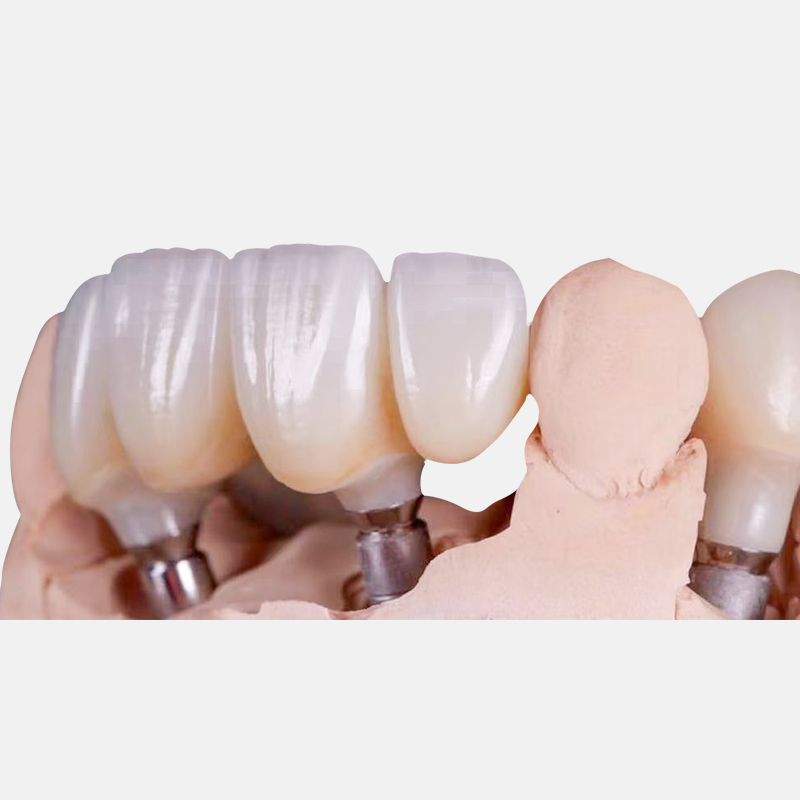
Capturing the precise metrics outlined above requires robust technology. We must evaluate three primary solution categories. Each carries unique advantages and distinct limitations depending on your clinical scenario.
Intraoral Scanning (IOS) with Scan Bodies: IOS technology is incredibly reliable for single units and short-span bridges. However, it struggles in full-arch scenarios featuring steeply angled implants. Cross-arch distortion often occurs. Software "stitching" errors frequently misalign the true trajectory of posterior tilted implants.
Photogrammetry: This technology is rapidly emerging as the gold standard for full-arch data capture. It calculates implant positions and angulations using dedicated extraoral cameras. The result delivers near-zero cross-arch distortion. It isolates the exact implant position independent of soft tissue interference.
Analog Impressions (Verification Jigs): The traditional baseline remains highly accurate. Splinting impression copings creates a rigid, reliable master model. Unfortunately, this analog workflow is labor-intensive. It also proves quite uncomfortable for the patient compared to digital alternatives.
Workflow Evaluation Dimensions Chart
Workflow Category | Scalability & Chair Time | Accuracy Validation | Ideal Clinical Application |
|---|---|---|---|
Intraoral Scanning (IOS) | Fast chairside capture, rapid digital processing | Software alignment relies heavily on scan body geometry | Short-span bridges, minor angulations |
Photogrammetry | Very fast chairside capture, automated processing | Direct mathematical calculation of 3D positions | Full-arch restorations, extreme posterior angulations |
Analog Impressions | Slow chairside procedure, requires physical shipping | Physical try-in validates the master cast perfectly | Complex cases showing digital inconsistency |
You must carefully weigh these evaluation dimensions. Consider scalability first. Time spent in the chair heavily influences patient satisfaction. Compare it against digital processing time in the laboratory. Accuracy validation remains paramount across any workflow. How accurately does the software align your angulation data alongside the soft tissue scan?
Keep skeptical considerations in mind regarding digital dentistry. No digital system guarantees perfect results automatically. Software interpolation errors frequently occur. If you seat scan bodies improperly, the data fails. If blood or saliva obscures the clinical field, scanners will guess the remaining geometry. You must verify every digital capture before authorizing fabrication.
The data you capture directly dictates the hardware you select. You need a reliable decision framework to navigate this process. Let us examine the two primary approaches for navigating challenging angulations.
This system operates as the workhorse for complex rehabilitations. It is best suited for implants placed at angles exceeding 15 to 30 degrees. MUAs correct the severe angulation at the tissue level. They standardize the restorative platform across the entire arch. This standardization creates a common path of insertion for the rigid framework. It is an absolutely essential component for full-arch cases. When utilizing this system, the multi unit abutment bridge distributes stress effectively away from the implant apex. It transforms divergent trajectories into parallel restorative platforms.
This approach works best for shallow implant angles. It is also ideal for highly specific aesthetic demands located in the anterior zone. A custom abutment allows precise control over the subgingival emergence profile. It supports the interproximal papilla perfectly. However, it complicates the path of insertion significantly. If multiple implants remain non-parallel, seating a rigid custom abutment bridge becomes mathematically impossible without generating extreme internal friction.
Seamless data integration ties these decisions together. The laboratory requires specific implant library files. They use these digital files to virtually align your chosen abutment type. The CAD software merges this library data with your scanned angulation data. If the initial data capture was flawed, the virtual alignment will generate a non-passive framework, regardless of the abutment system you choose.
An evidence-oriented risk assessment reveals clear clinical dangers. Steep implant angulations inherently increase bending moments. They introduce destructive off-axis loading forces during heavy mastication. If you ignore these biomechanical realities, the restoration will eventually fracture. The abutment screws will loosen repeatedly over time.
Material limitations play a massive role in risk mitigation. Angulation data often reveals inadequate restorative space over a tilted posterior implant. When this occurs, you must pivot your strategy. Certain materials simply cannot survive thin cross-sections. You must discard unreinforced acrylic in favor of stronger alternatives. Milled titanium bars or monolithic zirconia frameworks become mandatory. They possess the tensile strength needed to withstand applied leverage across steep angles.
Managing screw channel emergence presents another major clinical hurdle. You can utilize dynamic or angled screw channel (ASC) solutions. These systems allow up to 25 or 30-degree corrections inside the restoration itself. However, this approach requires specific laboratory software capabilities. You will also need proprietary screwdrivers to torque the final prosthesis accurately. Using standard drivers on ASC channels will strip the screw heads instantly.
We must emphasize the strict verification requirement. You should always mandate a printed try-in or a physical verification jig. Do this before finalizing the definitive prosthesis. This critical step validates the digital angulation data inside the patient's mouth. It represents the ultimate safety net against digital distortion. Skipping this step invites catastrophic misfits during the final delivery appointment.
Capturing and transferring precise angulation data serves as the true linchpin of passive fit. It ensures long-term biomechanical stability in all complex implant bridge designs. Without reliable metrics, you gamble on the longevity of the final restoration.
Consider these actionable next steps to improve your outcomes:
Audit your current impression or scanning workflow. Track your cross-arch deviation rates carefully over your next ten full-arch cases.
Evaluate your laboratory partners based on their advanced digital capabilities. Look for proven photogrammetry integration and dynamic screw channel design expertise.
Establish a standard operating procedure (SOP) for your clinic. Record and verify A-P spread and trajectory data physically prior to authorizing the final restorative phase.
Transition strictly to milled titanium or monolithic zirconia frameworks when managing severely tilted posterior implants to prevent structural fatigue.
A: While implants can be placed at 30 to 45 degrees to avoid anatomical structures, restorative correction typically caps at 30 degrees using angled MUAs or dynamic screw channels. Exceeding this limit dramatically increases the risk of mechanical complications and screw loosening.
A: For single units or short spans, yes. For full-arch cases, steep angles increase the risk of stitching errors. Supplementary data like photogrammetry or physical verification jigs are highly recommended to prevent cross-arch distortion.
A: Off-axis loading increases strain on the crestal bone and restorative screws. Accurate bridge design uses rigid frameworks and specific abutments to redirect these forces axially as much as possible, protecting the underlying osseous integration.


